How the Eye Works

The anatomy of the human eye and the way it works border on the miraculous. “How does the eye work?” is a formidable question, a detailed answer to which could easily fill a good-sized book, so what follows is necessarily a very brief overview.
When you look at a close or distant object, the light emitted from the object enters your eye and is processed in several stages. Finally it becomes a sharply focussed image on the retina (the back covering of the eye). Millions of tiny receptors in the retina change the image into electrical impulses and transmit them along the optic nerve to the brain, which either has the stored information for you to recognise the object or, if it hasn’t, will make you wonder what the object is.
It’s easier to understand how the eye works by identifying its main parts and their functions in the order in which light reaches them:
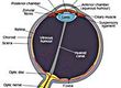
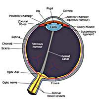
The Cornea
The cornea is the eye’s transparent, dome-shaped front covering. It is comparable to the lens in a basic camera, refracting (bending) incoming light rays to form an image on the retina. The cornea comprises about one thirteenth of the surface area of the entire eyeball (often called the globe).The Aqueous Humour
The anterior (front) chamber lies between the cornea and the iris and is filled with a transparent fluid, about 270 microlitres in volume, called the aqueous humour. The aqueous fluid slowly circulates in this chamber and around 1% of it is removed from the eye every minute to flow over the cornea. This process keeps the corneal cells refreshed throughout their life cycle (about 7 days) and carries away spent cells. There is some pressure in the aqueous which also helps to maintain the shape of the globe.The Iris
The iris is the opaque, round, coloured part of the eye surrounding the pupil, which is the opening that allows light into the back of the eye. Pigmentation in the iris is what defines the perceived colour of the eye. The action of the iris widens or shrinks the pupil, controlling the amount of light entering the eye according to the brightness of the object in view. The variable aperture on a camera (also called the iris) does the same job, the size of the aperture being controlled by a light meter.The Crystalline Lens
The crystalline lens lies immediately behind the iris. This lens is equivalent to the focussing mechanism in the camera. It is attached to the wall of the eye near the outer edges of the cornea by the surrounding ciliary muscles. These muscles expand or contract to change the shape of the lens to ‘accommodate’ for objects at varying distances. When the ciliary muscles are relaxed the eye is focussed for long distance. To view closer objects the ciliary muscles contract and the lens changes shape to ensure that the image is correctly focussed on the retina. In adults the diameter of the crystalline lens is about 9.5mm.The Vitreous Humour
The vitreous humour is a transparent gel that fills the eye (a volume of about 4 millilitres) between the crystalline lens to the retina.The Retina
This light-sensitive structure lines the inside of the back of the eye and can be likened to the film in a camera. The retina has eight separate layers, one of which is packed with receptors known as ‘rods and cones’. There are about 120 million rods and 7 million cones in the retina (with 127 megapixels to measure up to, the digital camera still has some way to go!). Rods are by far the most sensitive receptors and the most efficient at adapting to the dark, but they are not sensitive to colour. The cones do the colour work and they are way out in front when it comes to high-resolution vision. For this reason the concentration of cones is greatest at the centre of the retina in the area known as the fovea and the eye moves to keep the image in the foveal area.Another layer of the retina houses a network of nerve fibres that carry electrical impulses from the rods and cones to the head of the optic nerve, which is on the retina about 4mm from its centre, and then to the brain.
The human eye certainly is an amazing organ. If you were building a robot, you would have one or two problems copying the eye, although technology is always on the case and is getting closer all the time. The digital camera and light-sensitive and passive infra-red switches are modern examples of the progress. Medical science has also recently succeeded in developing an implant containing ‘pixels’ that replace missing or damaged receptors in the retina and connect to the optic nerve.
Keep an eye on the progress.